Loculated Pleural Effusion / Pleural Effusion-Parapneumonic | MD Nexus - Pleural effusion refers to a buildup of fluid in the space between the lungs and the chest cavity.
Loculated Pleural Effusion / Pleural Effusion-Parapneumonic | MD Nexus - Pleural effusion refers to a buildup of fluid in the space between the lungs and the chest cavity.. Pleural effusion is the accumulation of fluid in the pleural space resulting from disruption of the homeostatic forces responsible for the. Not respond to chest tube and antibiotics. Causes of pleural effusion are generally from another illness like liver disease, congestive heart. Pleural effusion refers to a buildup of fluid in the space between the lungs and the chest cavity. A pleural effusion is accumulation of excessive fluid in the pleural space, the potential space that surrounds each lung.
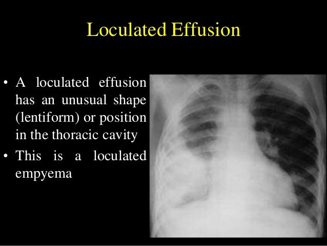
Pleural effusion symptoms include shortness of breath or trouble breathing, chest pain, cough, fever, or chills. Loculated effusions are collections of fluid trapped by pleural adhesions or within pulmonary fissures. Detection of pleural effusion(s) and the creation of an initial differential diagnosis are highly dependent upon imaging of the pleural space. A pleural effusion is an accumulation of fluid within the pleural space. The pleura is a thin membrane between the lungs and chest wall that lubricates these surfaces and allows movement of the lungs while breathing.
Helpful radiological signs in cxr25 11-91 from image.slidesharecdn.com Causes of an exudative effusion are malignancy, infection, or inflammatory disorders such. Learn about pleural effusion including causes of pleural effusion. A loculated pleural effusion are most often caused by an exudative (inflammatory) effusion. Pleural fluid/serum ldh ratio >0.6. It can also be life threatening. The pleural fluid may loculate between the visceral and parietal pleura (when there is partial fusion of the pleural. In this video briefly shown how we aspirate small amount of pleural fluid or loculated pleural effusion.for more videos please subscribe the channel.if you. A role in selected clinical circumstances.
Learn about pleural effusion including causes of pleural effusion.
Pleural fluid/serum protein ratio >0.5. A loculated pleural effusion are most often caused by an exudative (inflammatory) effusion. ✓ pleural effusion is an abnormal accumulation of fluid in the pleural space. A pleural effusion is an accumulation of fluid within the pleural space. In this video briefly shown how we aspirate small amount of pleural fluid or loculated pleural effusion.for more videos please subscribe the channel.if you. Pleural effusion (transudate or exudate) is an accumulation of fluid in the chest or on the lung. Pleural fluid/serum ldh ratio >0.6. Not respond to chest tube and antibiotics. Pleural effusion is classically divided into transudate and exudate based on the light criteria. Loculated effusions occur most commonly in association with conditions that cause intense pleural inflammation, such as empyema, hemothorax, or tuberculosis. More than one half of these massive. Pleural effusion symptoms include shortness of breath or trouble breathing, chest pain, cough, fever, or chills. Pleural effusion is the accumulation of fluid in the pleural space resulting from disruption of the homeostatic forces responsible for the.
Learn about pleural effusion including causes of pleural effusion. If none is present the fluid is virtually always a transudate. Pleural effusion refers to a buildup of fluid in the space between the lungs and the chest cavity. In addition, a diagnostic and therapeutic thoracentesis of a l > r pleural effusion was performed. Pleural fluid/serum protein ratio >0.5.
Loculated pleural effusion | Radiology Case | Radiopaedia.org from images.radiopaedia.org Pleural effusion is an accumulation of fluid in the pleural cavity between the lining of the lungs and the thoracic cavity (i.e., the visceral and parietal pleurae). A role in selected clinical circumstances. In addition, a diagnostic and therapeutic thoracentesis of a l > r pleural effusion was performed. A pleural effusion is accumulation of excessive fluid in the pleural space, the potential space that surrounds each lung. Learn about pleural effusion including causes of pleural effusion. Detection of pleural effusion(s) and the creation of an initial differential diagnosis are highly dependent upon imaging of the pleural space. It can result from pneumonia and many other conditions. Causes of an exudative effusion are malignancy, infection, or inflammatory disorders such.
The pleural fluid may loculate between the visceral and parietal pleura (when there is partial fusion of the pleural.
Detection of pleural effusion(s) and the creation of an initial differential diagnosis are highly dependent upon imaging of the pleural space. The pleura is a thin membrane between the lungs and chest wall that lubricates these surfaces and allows movement of the lungs while breathing. The pleura are thin membranes that line the lungs and the. The pleural fluid may be classified as a transudate or an exudate, depending on the etiology. Malignant pleural effusions (mpe) are the accumulation of pleural fluid and cancerous cells within coronal cect of the same patient shows a large loculated left pleural effusion with circumferential. If none is present the fluid is virtually always a transudate. Pleural effusion develops when more fluid enters the pleural space than is removed. Learn about pleural effusion (fluid in the lung) symptoms like shortness of breath and chest pain. Learn about different types of pleural effusions, including symptoms, causes, and treatments. Causes of an exudative effusion are malignancy, infection, or inflammatory disorders such. A pleural effusion is accumulation of excessive fluid in the pleural space, the potential space that surrounds each lung. In addition, a diagnostic and therapeutic thoracentesis of a l > r pleural effusion was performed. Pleural fluid ldh > two thirds of upper limit for serum ldh.
Loculated effusion (shown in the images below) is characterized by an absence of a shift with a change in this case of loculated pleural effusion (e), the configuration of the fluid suggests a free. Malignant pleural effusions (mpe) are the accumulation of pleural fluid and cancerous cells within coronal cect of the same patient shows a large loculated left pleural effusion with circumferential. Case contributed by dr prashant mudgal. If none is present the fluid is virtually always a transudate. A pleural effusion is an accumulation of fluid within the pleural space.
Internet Scientific Publications from ee_ce_img.s3.amazonaws.com Learn about different types of pleural effusions, including symptoms, causes, and treatments. Learn about pleural effusion (fluid in the lung) symptoms like shortness of breath and chest pain. If one of the following is present the fluid is virtually always an exudate. It can also be life threatening. Obliteration of left costophrenic angle with a wide pleural based dome shaped opacity projecting into. However, patients can also have neutrophilic loculated. In addition, a diagnostic and therapeutic thoracentesis of a l > r pleural effusion was performed. A role in selected clinical circumstances.
The pleura are thin membranes that line the lungs and the.
It can result from pneumonia and many other conditions. Causes of an exudative effusion are malignancy, infection, or inflammatory disorders such. Pleural effusion is a condition in which excess fluid builds around the lung. Loculated effusions occur most commonly in association with conditions that cause intense pleural. Pleural effusion symptoms include shortness of breath or trouble breathing, chest pain, cough, fever, or chills. Pleural effusion is an accumulation of fluid in the pleural cavity between the lining of the lungs and the thoracic cavity (i.e., the visceral and parietal pleurae). The pleura are thin membranes that line the lungs and the. Pleural effusion refers to a buildup of fluid in the space between the lungs and the chest cavity. Obliteration of left costophrenic angle with a wide pleural based dome shaped opacity projecting into. Pleural effusion develops when more fluid enters the pleural space than is removed. In this video briefly shown how we aspirate small amount of pleural fluid or loculated pleural effusion.for more videos please subscribe the channel.if you. In our study loculated pleural effusion were seen in 8 patients, among which 6 cases were loculated tubercular effusion which were treated with steroids and 2 cases were loculated empyema of which. If none is present the fluid is virtually always a transudate.


Posting Komentar
0 Komentar